Mammogram breast cancer screening
A mammogram is an X-ray image of the breast used as a screening tool to detect early signs of breast cancer, often before symptoms appear.
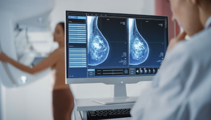
It plays a critical role in reducing breast cancer mortality by allowing for early diagnosis, when treatment is most effective. It is a simple outpatient test, done in few minutes with minimal discomfort but goes a very long way in identifying cancer very early.
Key Features of Nipple-Sparing Mastectomy

- Early Detection: Mammograms can detect tumors that are too small to be felt, as well as microcalcifications (tiny calcium deposits) that may indicate early-stage breast cancer.
- Reducing Mortality: Detecting breast cancer at an early stage improves survival rates and often allows for less aggressive treatment options.
- Tracking Changes: Mammograms provide a baseline image of the breast, which allows doctors to compare changes over time. This is useful for monitoring abnormalities. Certain changes seen on mammogram may not be obvious malignancy and in such situations mammograms done over years are compared to look for changes
- Screening Mammograms: Performed on women who do not have symptoms of breast cancer, typically to detect cancer early. The screening mammogram usually involves two X-ray images of each breast (top-to-bottom and side-to-side views).
- Diagnostic Mammograms: Performed when there are signs or symptoms of breast cancer (e.g., a lump or unusual discharge), or if something suspicious is found on a screening mammogram. Diagnostic mammograms provide more detailed images to investigate potential abnormalities.
- Positioning: The breast is placed on a flat surface, and a plastic plate compresses the breast tissue to spread it out. This compression ensures that the X-ray can capture a clear image with minimal overlap of tissue.
- X-ray Images: X-rays are taken from different angles. The compression may feel uncomfortable, but it only lasts for a few seconds.
- Duration: The whole process usually takes about 20 minutes, with the actual compression lasting just a few seconds for each image.
- Results: A radiologist analyzes the images for any signs of abnormalities, such as masses, asymmetries, or calcifications.
- General Guidelines:
- Women aged 40 to 44: Option to start annual mammograms if they wish.
- Women aged 45 to 54: Recommended to have a mammogram every year.
- Women aged 55 and older: Can switch to mammograms every two years or continue yearly screening, depending on health status and personal choice.
- High-Risk Women: Women with a higher risk of breast cancer (e.g., those with a family history of breast cancer or BRCA1/BRCA2 gene mutations) may need to begin screening earlier and may also need additional imaging tests, such as breast MRI, along with mammograms.
- Older Women: Screening may continue for women over 75, depending on their health and life expectancy.
- Reduction in Mortality: Regular mammograms can reduce the risk of dying from breast cancer by detecting cancer before it spreads.
- Detects Cancer Early: Tumors found through mammography are typically smaller and more treatable.
- Non-invasive: It is a relatively simple and non-invasive procedure.
- What it is: 3D mammography takes multiple images of the breast from different angles and creates a three-dimensional picture. It is often used in combination with traditional 2D mammography.
- Advantages: 3D mammograms are particularly useful for women with dense breasts or those at higher risk of breast cancer, as they provide clearer images and can reduce the likelihood of false positives.
- Improved Accuracy: Studies suggest that 3D mammography may detect more cancers and reduce the need for follow-up testing.
- Normal Results: If no abnormalities are found, you’ll be advised when to schedule your next screening.
- Abnormal Results: If something suspicious is found, the radiologist may recommend further tests, such as a diagnostic mammogram, ultrasound, or biopsy, to determine if the abnormality is cancerous.
- What are Dense Breasts?: Breasts with more fibrous and glandular tissue than fatty tissue. Dense tissue appears white on a mammogram, which can make it more difficult to see tumors (which also appear white).
- Supplemental Screening: Women with dense breasts may benefit from additional imaging, such as ultrasound or MRI, to improve the chances of detecting cancer.
- Sensitivity: The ability of the mammogram to correctly identify cancer depends on several factors, including the patient’s age, breast density, and overall risk factors.
- Higher sensitivity: Mammograms tend to be more accurate for older women, particularly those over 50, as their breasts tend to be less dense.
- Lower sensitivity: For women with dense breasts or younger women, the mammogram may be less sensitive and can sometimes miss cancers.
- Lower sensitivity: For women with dense breasts or younger women, the mammogram may be less sensitive and can sometimes miss cancers. • Improving Accuracy: To improve the detection rate, doctors may recommend other imaging techniques like 3D mammography, ultrasound, or MRI, particularly for high-risk individuals or those with dense breasts.
Mammogram is always done along with breast ultrasound, as certain changes in the breast may not be visible on mammogram and such changes can be picked up on the breast ultrasound. Further it is a myth, taking mammogram gives lot of radiation to the body and mammogram is very safe and effective test for breast cancer screening.
